Presentation
The Institutional Microscopy Laboratory has 4 microscopes that can be used in diferent techniques.
Equipment
Details about the equipment that can be found in the laboratory.
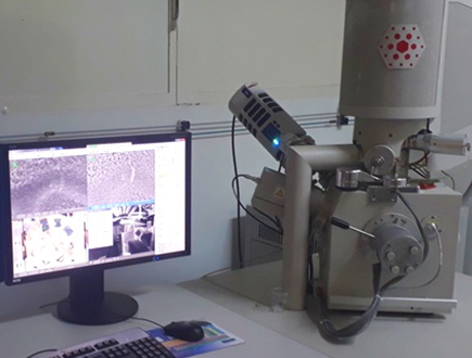
Scanning Electron Microscope - FEI Quanta FEG 250
The Quanta FEG-250 is a field emission scanning electron microscope (FEG-SEM), designed for analyses requiring high resolution and for samples sensitive to the electron beam, such as polymers, natural fibers, and cellulose. It operates with excellent performance even at low accelerating voltages (≈2 kV), enabling the characterization of nanometric materials such as graphene oxide and cellulose nanocrystals with minimal sample degradation.
It is equipped with the following detectors:
• Everhart-Thornley Detector (ETD): Secondary electron detector used for topographic imaging.
• Circular Backscatter Detector (CBS): Backscattered electron detector that provides both topographic images and atomic number contrast images.
• Large Field Detector (LFD): Used in Low Vacuum mode to obtain images of non-conductive or non-metalized samples.
•Environmental Scanning Electron Microscopy (ESEM): Enables imaging in “Environmental Mode,” a low-vacuum mode suitable for samples with moderate water content (20–30%) or under humid conditions.
• Scanning Transmission Electron Microscopy (STEM): Provides high-resolution imaging in transmission mode with a focused electron beam (magnifications up to ~300,000x). Ideal for nanostructured samples.
• Energy Dispersive Spectrometer (EDS – Oxford X-Max 50): Allows semi-quantitative elemental analyses (spot, line scan, or mapping) with high sensitivity and spatial resolution.
Samples not recommended for analysis:
• Containing volatile substances
• With high water content
• Containing solvent/oil
• Magnetic
• Corrosive
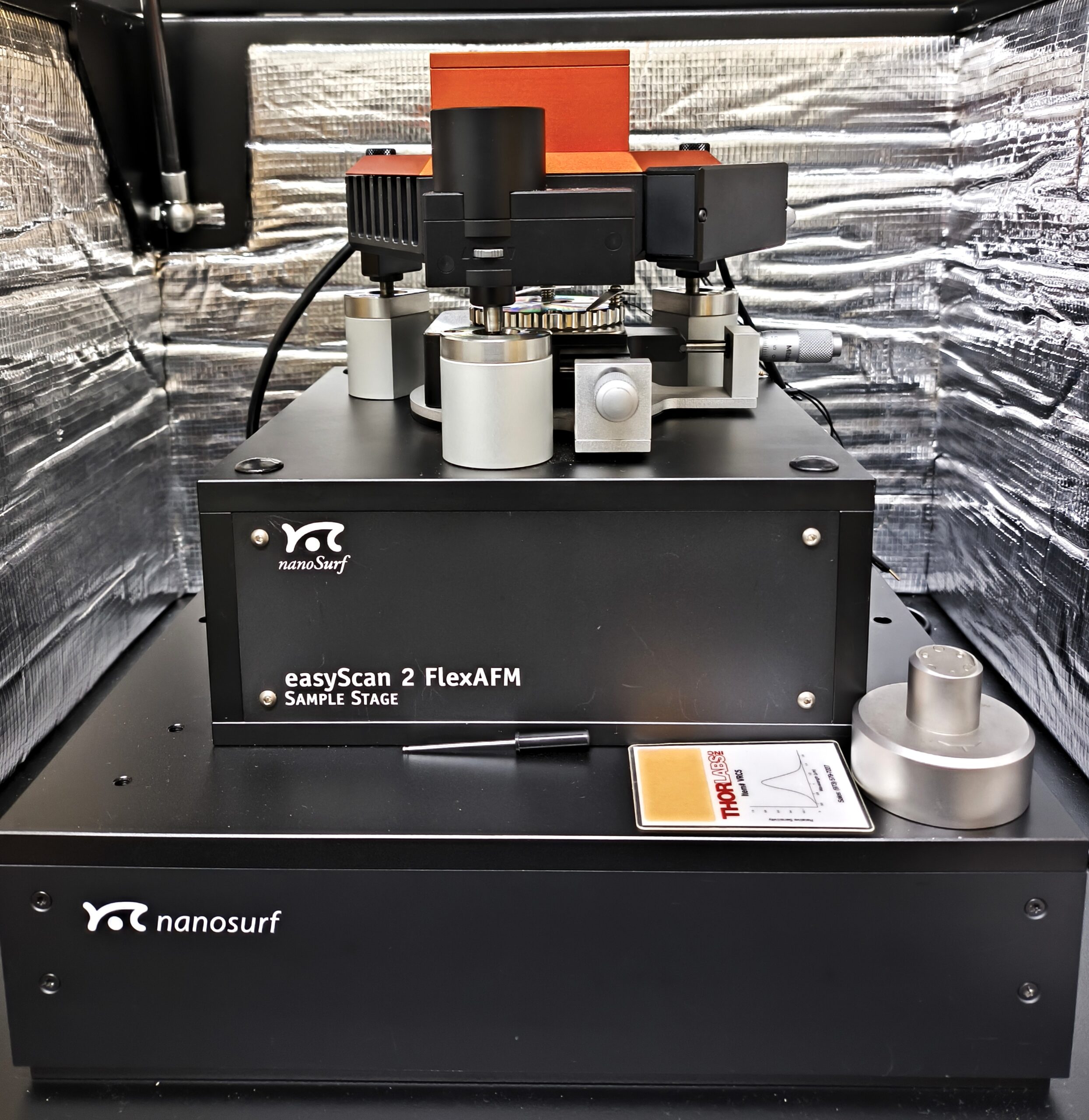
Atomic Force Microscope - Nanosurf C3000 FlexAFM
The Atomic Force Microscope (AFM) enables analyses under ambient conditions with minimal sample preparation, serving as a complementary technique to Scanning Electron Microscopy (SEM). The FlexAFM model with C3000 controller has an x-y scan range of 100 μm and a z-axis range of 10 μm.
It is suitable for surface analysis of a wide range of materials, including those that cannot be analyzed by SEM, such as:
• Materials with high water content
• Materials containing solvent and/or oil residues, such as petroleum and asphaltenes
• Magnetic materials
The main operation modes are as follows:
• Non-contact/tapping: standard mode of operation for AFM, that enables acquisition of topography and phase contrast.
• Kelvin Probe Force Microscopy (KPFM): allows simultaneous mapping of surface potential and topography.
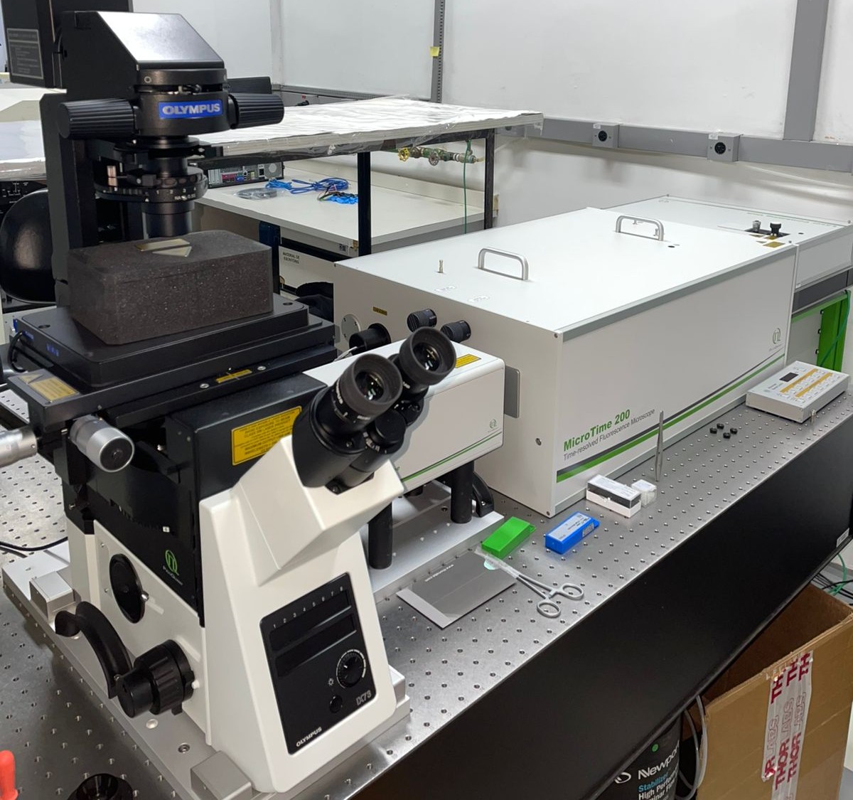
Scanning Confocal Microscope – PicoQuant MicroTime 200
The MicroTime 200 is a highly sensitive confocal fluorescence microscope designed for single-molecule studies and quantitative analysis of dynamic processes in biological, chemical, and materials science systems. Its flexible and open design allows both routine experiments and adaptation to highly specialized research projects.
It is equipped with the following modes and analysis techniques:
• Fluorescence Correlation Spectroscopy (FCS) – determines molecular mobility, diffusion coefficients, and concentrations in solutions or living cells.
• Fluorescence Lifetime Imaging (FLIM) – discriminates spectrally similar fluorophores, analyzes molecular interactions, and eliminates background autofluorescence.
• Förster Resonance Energy Transfer (FRET) – measures inter- and intramolecular distances, detects interactions, and monitors conformational changes of biomolecules.
• Single-molecule measurements – enable observation of processes such as aggregation, association/dissociation, and photophysics of individual fluorophores.
• Fluorescence Intensity Time Traces – monitor blinking of single particles, photobleaching, and fluorophore stability.
The system is equipped with excitation lasers at 405, 440, 470, 510, and 640 nm, and two detectors (SPAD and Hybrid PMT) that allow simultaneous signal acquisition in two channels.
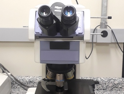
Optical Microscope – Nikon E800

Sample Preparation Equipment: Metalizer – Baltec MED020
The metal coater is essential for sample preparation in scanning electron microscopy, applying coatings that prevent charge buildup on non-conductive materials.
The equipment allows coatings by:
• Sputtering (with Au/Pd alloy or metallic Ir);
• Carbon evaporation.
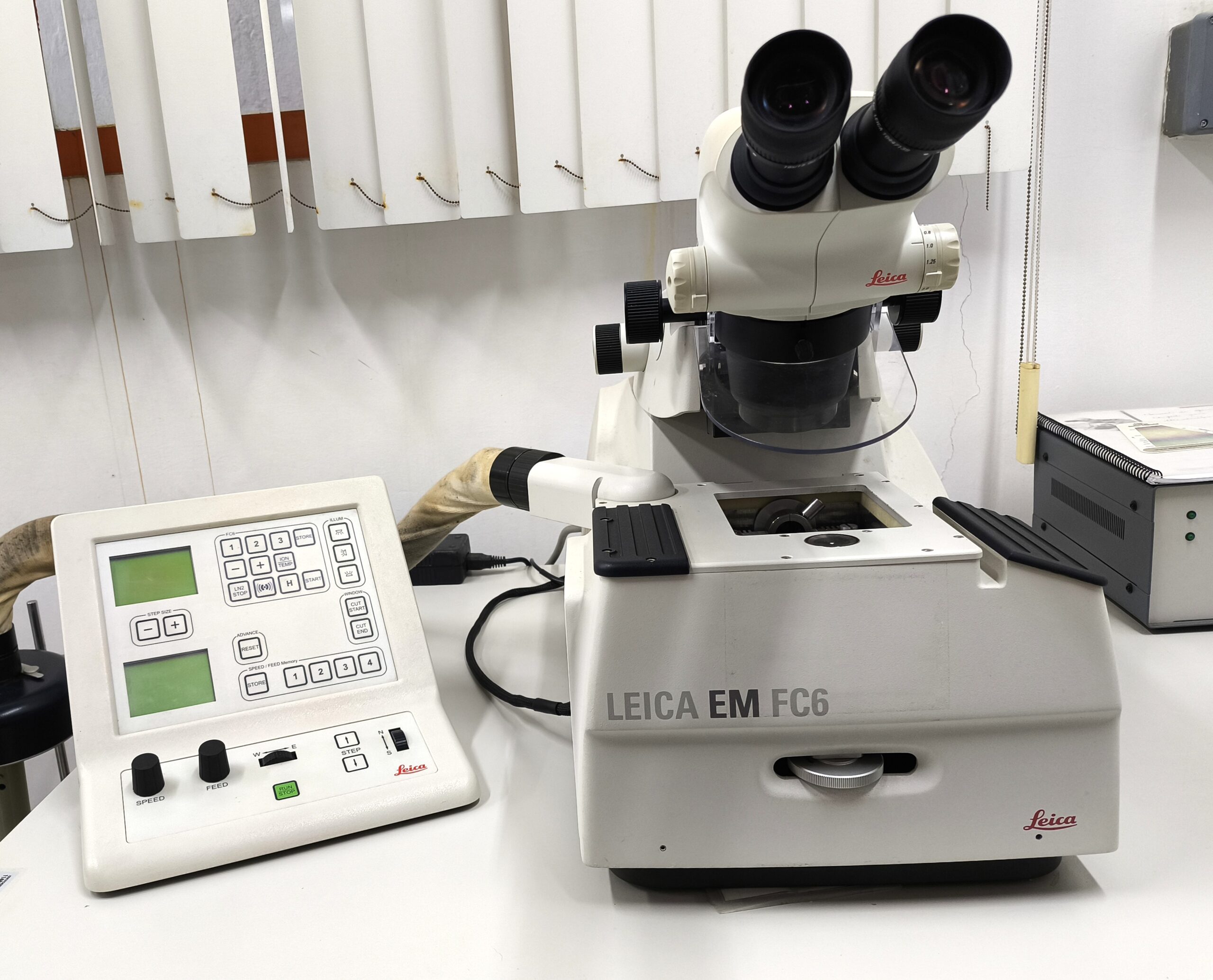
Sample Preparation Equipment: Ultra Cryomicrotome – Leica FC6
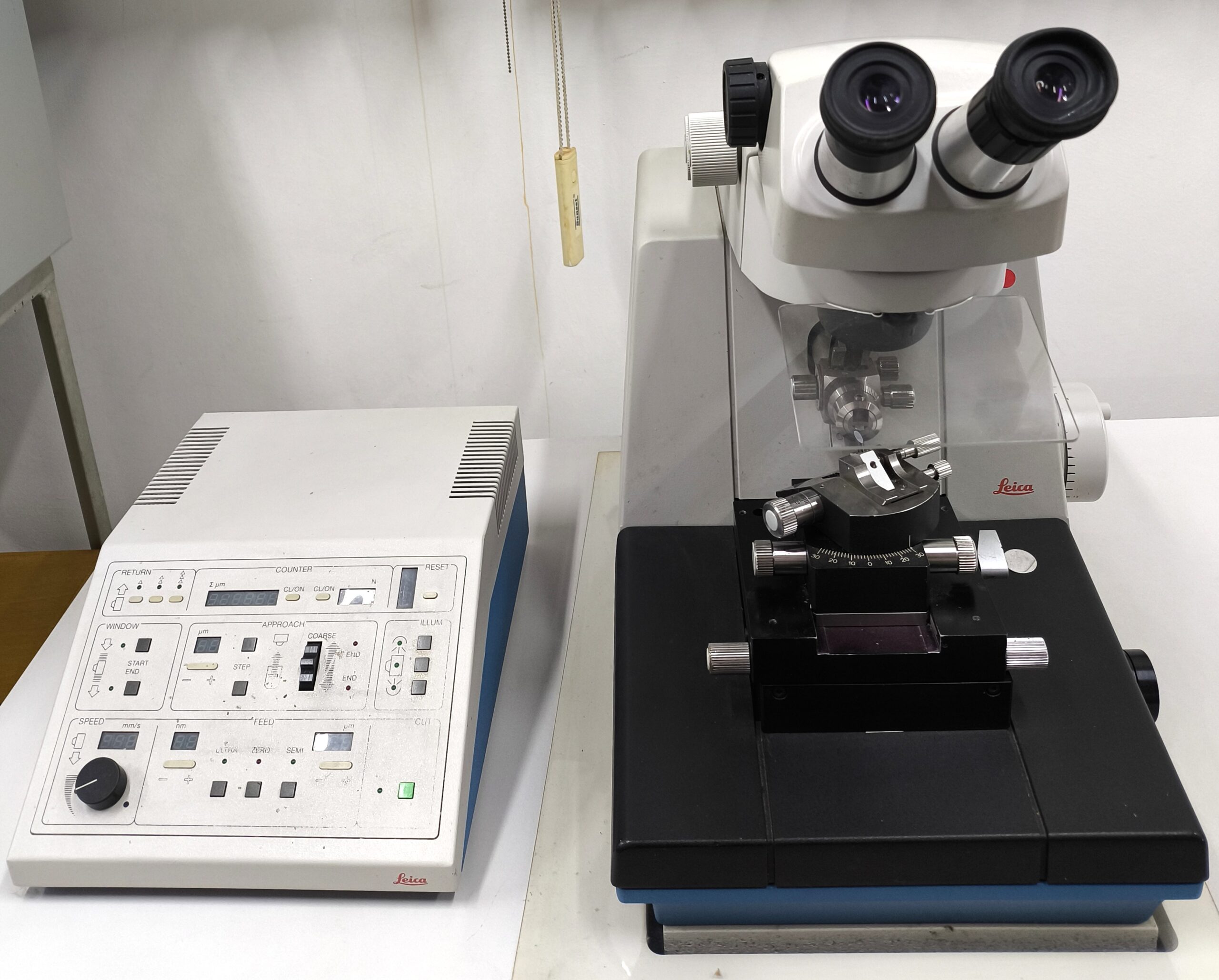
Sample Preparation Equipment: Ultramicrotome – Leica ULTRACUT S
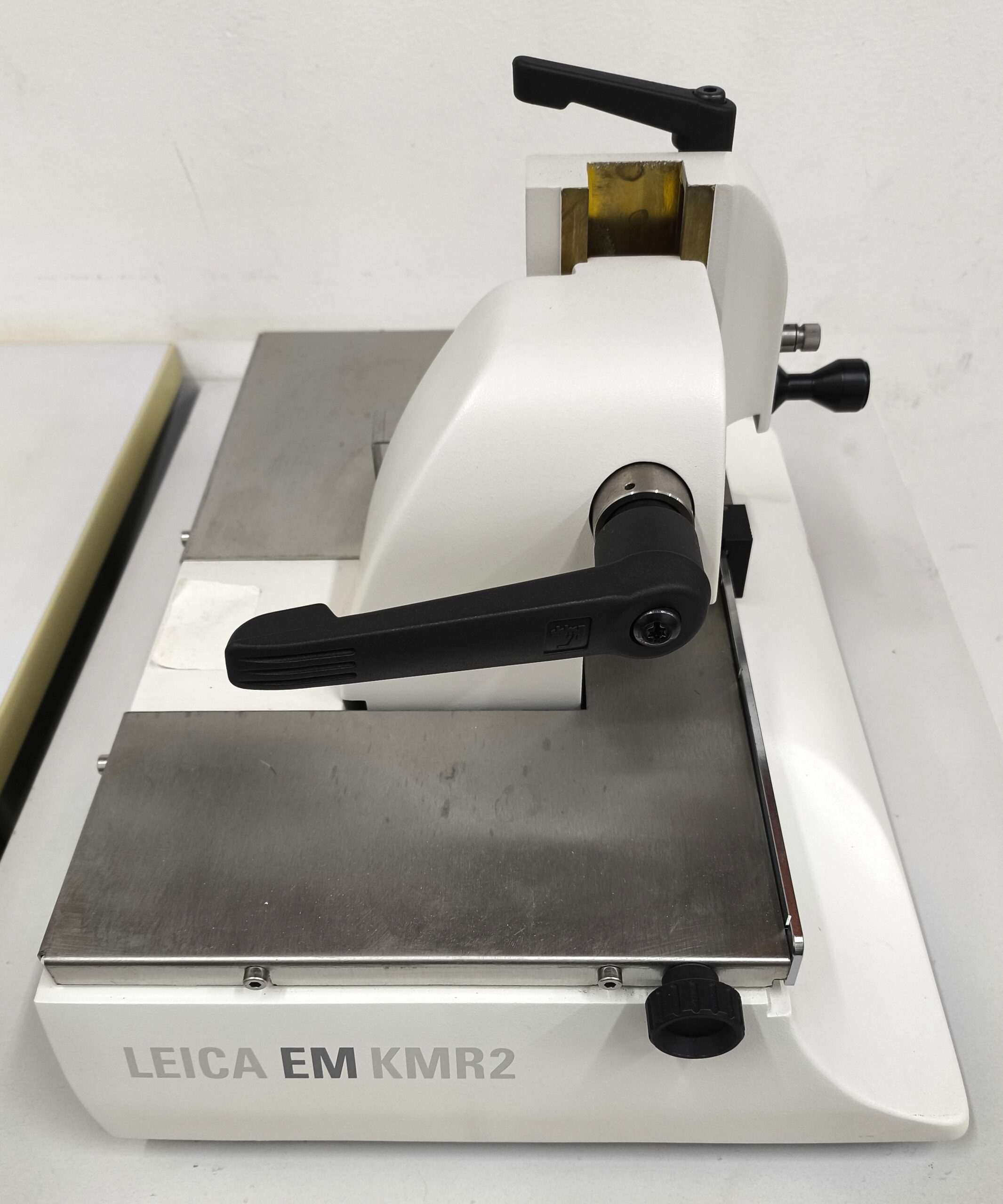
Sample Preparation Equipment: Device for Fabrication of Triangular Glass Knives – Leica KMR2
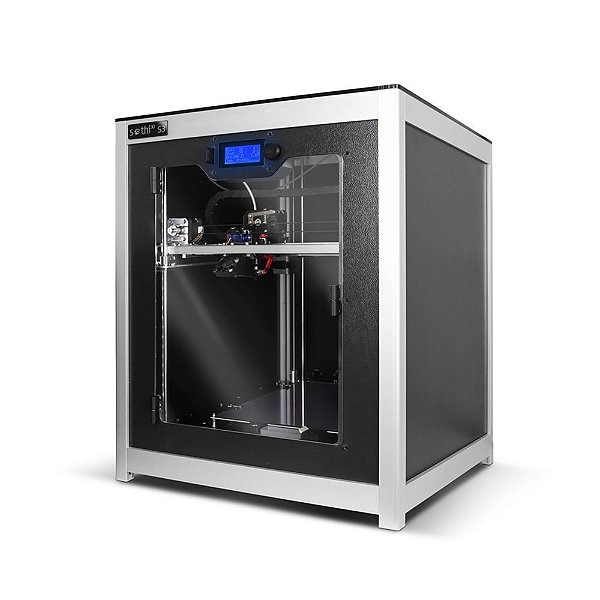
Printer – FDM Sethi3D S3
Printing area: 270 × 270 × 320 mm (23.3 liters)
Extruder: 0.2 mm nozzle
Filament: 1.75 mm
Layer height: adjustable from 50 μm (0.05 mm) to 300 μm (0.3 mm)
Printing speed: up to 150 mm/s;
travel speed: up to 300 mm/s
Heated aluminum bed
Computer connection: USB interface
Location: Lab B-207
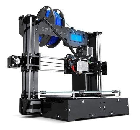
Printer – FDM Sethi3D AiP
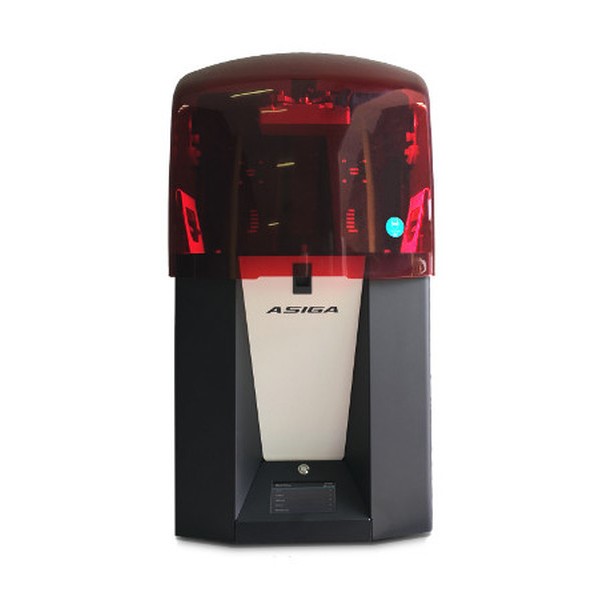
Printer – DLP ASIGA FreeForm UV-Pro2
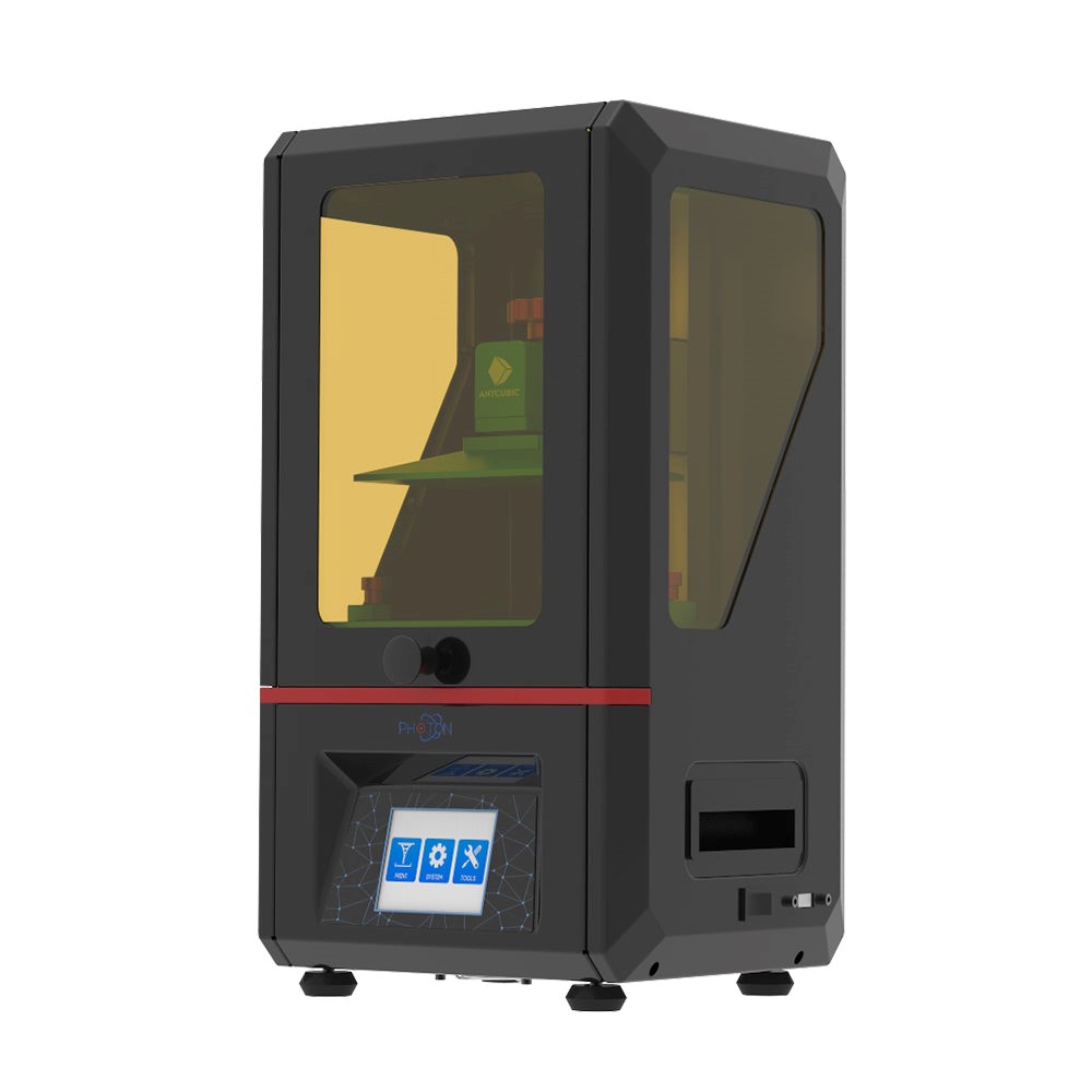
Printer – LCD Anycubic Photon 2K
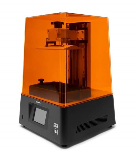
Printer – LCD Phrozen Sonic Mini 8K
Printer – LCD Anycibic Photon Mono M5s
Team

Hugo Campos Loureiro, M. Sc.